|
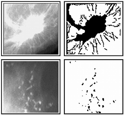
The aim of the segmentation step in mammographic image analysis is to extract regions of interest (ROIs) containing all breast abnormalities from the normal breast tissue. Another aim of the segmentation is to locate the suspicious lesion candidates from the region of interest. H.D. Li, M. Kallergi, L.P. Clarke, V.K. Jain, R.A. Clark, Markov Random Field for Tumor Detection in Digital Mammography, IEEE Transactions on Medical Imaging, Vol. 14, No. 3, 1995, pp. 565-576 F. Lefebvre, H. Benali, R. Gilles, E. Kahn, R. Di Paola, A Fractal Approach to the Segmentation of Microcalcifications in Digital Mammograms, Medical Physics, Vol. 22, No. 4, 1995, pp. 381-390 W. Qian, M. Kallergi, L.P. Clarke, H.-D. Li, P. Venugopal, D. Song, R.A. Clark, Tree Structured Wavelet Transform Segmentation of Microcalcifications in Digital Mammography, Medical Physics, Vol. 22, No. 8, 1995, pp. 1247-1254 N. Patrick, H.-P. Chan, B. Sahiner, D. Wei, An Adaptive Density-Weighted Contrast Enhancement Filter for Mammographic Breast Mass Detection, IEEE Transactions on Medical Imaging, Vol. 15, No. 1, 1996, pp. 59-67 H. Li, K.J.R. Liu, S.-C.B. Lo, Fractal Modeling and Segmentation for the Enhancement of Microcalcifications in Digital Mammograms, IEEE Transactions on Medical Imaging, Vol. 16, No. 6, 1997, pp. 785-798 M.A. Kupinski, M.L. Giger, Automated Seeded Lesion Segmentation on Digital Mammograms, IEEE Transactions on Medical Imaging, Vol. 17, No. 4, 1998, pp. 510-517 N. Petrick, H.-P. Chan, B. Sahiner, M.A. Helvie, Combined Adaptive Enhancement and Region-Growing Segmentation of Breast Masses on Digitized Mammograms, Medical Physics, Vol. 26, No. 8, 1999, pp. 1642-1654 M.A. Gavrielides, J.Y. Lo, R. Vargas-Voracek, C.E. Floyd Jr., Segmentation of Suspicious Clustered Microcalcifications in Mammograms, Medical Physics, Vol. 27, No. 1, 2000, pp. 13-22 G.M. te Brake, N. Karssemeijer, Segmentation of Suspicious Densities in Digital Mammograms, Medical Physics, Vol. 28, No. 2, 2001, pp. 259-266 H. Li, Y. Wang, K.J.R. Liu, S.-C.B. Lo, M.T. Freedman, Computerized Radiographic Mass Detection - Part I: Lesion Site Selection by Morphological Enhancement and Contextual Segmentation, IEEE Transactions on Medical Imaging, Vol. 20, No. 4, 2001, pp. 289-301 A.R. Domínguez, A.K. Nandi, Improved Dynamic-Programming-Based Algorithms for Segmentation of Masses in Mammograms, Medical Physics, Vol. 34, No. 11, 2007, pp. 4256-4269 back to top
|
 |
|
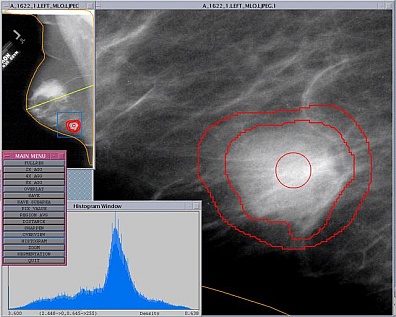
In the feature extraction step of the mammographic image analysis algorithms the features are calculated from the characteristics of the region of interest. C.J. D'Orsi, D.B. Kopans, Mammographic Feature Analysis, Seminars in Roentgenology, Vol. 28, No. 3, 1993, pp. 204-230 Z. Huo, M.L. Giger, C.J. Vyborny, U. Bick, P. Lu, D.E. Wolverton, R.A. Schmidt, Analysis of Spiculation in the Computerized Classification of Mammographic Masses, Medical Physics, Vol. 22, No. 10, 1995, pp. 1569-1579 A.P. Dhawan, Y. Chitre, C. Kaiser-Bonasso, M. Moskowitz, Analysis of Mammographic Microcalcifications Using Gray-Level Image Structure Features, IEEE Transactions on Medical Imaging, Vol. 15, No. 3, 1996, pp. 246-259 B. Sahiner, H.-P. Chan, N. Petrick, M.A. Helvie, M.M. Goodsitt, Computerized Characterization of Masses on Mammograms: the Rubber Band Straightening Transform and Texture Analysis, Medical Physics, Vol. 25, No. 4, 1998, pp. 516-526 H.-P. Chan, B. Sahiner, K.L. Lam, N. Petrick, M.A. Helvie, M.M. Goodsitt, D.D. Adler, Computerized Analysis of Mammographic Microcalcifications in Morphological and Texture Feature Spaces, Medical Physics, Vol. 25, No. 10, 1998, pp. 2007-2019 W. Qian, L. Li, L.P. Clarke, Image Feature Extraction for Mass Detection in Digital Mammography: Influence of Wavelet Analysis, Medical Physics, Vol. 26, No. 3, 1999, pp. 402-408 H. Kobatake, M. Murakami, H. Takeo, S. Nawano, Computerized Detection of Malignant Tumors on Digital Mammograms, IEEE Transactions on Medical Imaging, Vol. 18, No. 5, 1999, pp. 369-378 N.R. Mudigonda, R.M. Rangayyan, J.E.L. Desautels, Gradient and Texture Analysis for the Classification of Mammographic Masses, IEEE Transactions on Medical Imaging, Vol. 19, No. 10, 2000, pp. 1032-1043 B. Verma, J. Zakos, A Computer-Aided Diagnosis System for Digital Mammograms Based on Fuzzy-Neural and Feature Extraction Techniques, IEEE Transactions on Information Technology in Biomedicine, Vol. 5, No. 1, 2001, pp. 46-54 L. Hadjiiski, B. Sahiner, H.-P. Chan, N. Petrick, M.A. Helvie, M. Gurcan, Analysis of Temporal Changes of Mammographic Features: Computer-Aided Classification of Malignant and Benign Breast Masses, Medical Physics, Vol. 28, No. 11, 2001, pp. 2309-2317 S. Timp, N. Karssemeijer, Interval Change Analysis to Improve Computer Aided Detection in Mammography, Medical Image Analysis, Vol. 10, No. 1, 2006, pp. 82-95 back to top
|
 |
|
Some of the features extracted from the regions of interest in the mammographic image are not significant when observed alone, but in combination with other features they can be significant for classification. The best set of features for eliminating false positives and for classifying lesion types as benign or malignant are selected in the feature selection step. B. Sahiner, H.-P. Chan, D. Wei, N. Petrick, M.A. Helvie, D.D. Adler, M.M. Goodsitt, Image Feature Selection by a Genetic Algorithm: Application to Classification of Mass and Normal Breast Tissue, Medical Physics, Vol. 23, No. 10, 1996, pp. 1671-1684 B. Zheng, Y.-H. Chang, X.-H. Wang, W.F. Good, D. Gur, Feature Selection for Computerized Mass Detection in Digitized Mammograms by Using a Genetic Algorithm, Academic Radiology, Vol. 6, No. 6, 1999, pp. 327-332 Z. Huo, M.L. Giger, D.E. Wolverton, W. Zhong, S. Cumming, O.I. Olopade, Computerized Analysis of Mammographic Parenchymal Patterns for Breast Cancer Risk Assessment: Feature Selection, Medical Physics, Vol. 27, No. 1, 2000, pp. 4-12 R.J. Nandi, A.K. Nandi, R.M. Rangayyan, D. Scutt, Classification of Breast Masses in Mammograms Using Genetic Programming and Feature Selection, Medical and Biological Engineering and Computing, Vol. 44, No. 8, 2006, pp. 683-694 B. Verma, P. Zhang, A Novel Neural-Genetic Algorithm to Find the Most Significant Combination of Features in Digital Mammograms, Applied Soft Computing Journal, Vol. 7, No. 2, 2007, pp. 612-625 back to top
|
 |
|
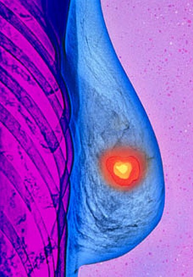
In the classification step of the mammographic image analysis algorithms lesions are classified as benign or malignant on the basis of selected features. Y. Wu, K. Doi, M.L. Giger, R.M. Nishikawa, Computerized Detection of Clustered Microcalcifications in Digital Mammograms: Applications of Artificial Neural Networks, Medical Physics, Vol. 19, No. 3, 1992, pp. 555-560 H.-P. Chan, D. Wei, M.A. Helvie, B. Sahiner, D.D. Adler, M.M. Goodsitt, N. Petrick, Computer-Aided Classification of Mammographic Masses and Normal Tissue: Linear Discriminant Analysis in Texture Feature Space, Physics in Medicine and Biology, Vol. 40, No. 5, 1995, pp. 857-876 H.-P. Chan, S.-C.B. Lo, B. Sahiner, Kwok Leung Lam, M.A. Helvie, Computer-Aided Detection of Mammographic Microcalcifications: Pattern Recognition with an Artificial Neural Network, Medical Physics, Vol. 22, No. 10, 1995, pp. 1555-1567 Y. Jiang, R.M. Nishikawa, D.E. Wolverton, C.E. Metz, M.L. Giger, R.A. Schmidt, C.J. Vyborny, K. Doi, Malignant and Benign Clustered Microcalcifications: Automated Feature Analysis and Classification, Radiology, Vol. 198, No. 3, 1996, pp. 671-678 B. Sahiner, H.-P. Chan, N. Petrick, D. Wei, M.A. Helvie, D.D. Adler, M.M. Goodsitt, Classification of Mass and Normal Breast Tissue: A Convolution Neural Network Classifier with Spatial Domain and Texture Images, IEEE Transactions on Medical Imaging, Vol. 15, No. 5, 1996, pp. 598-610 H.-P. Chan, B. Sahiner, N. Patrick, M.A. Helvie, K.L. Lam, D.D. Adler, M.M. Goodsitt, Computerized Classification of Malignant and Benign Microcalcifications on Mammograms: Texture Analysis Using an Artificial Neural Network, Physics in Medicine and Biology, Vol. 42, No. 3, 1997, pp. 549-567 Z. Huo, M.L. Giger, C.J. Vyborny, D.E. Wolverton, R.A. Schmidt, K. Doi, Automated Computerized Classification of Malignant and Benign Masses on Digitized Mammograms, Academic Radiology, Vol. 5, No. 3, 1998, pp. 155-168 W.J.H. Veldkamp, N. Karssemeijer, J.D.M. Otten, J.H.C.L. Hendriks, Automated Classification of Clustered Microcalcifications into Malignant and Benign Types, Medical Physics, Vol. 27, No. 11, 2000, pp. 2600-2608 A. Papadopoulos, D.I. Fotiadis, A. Likas, An Automatic Microcalcification Detection System Based on a Hybrid Neural Network Classifier, Artificial Intelligence in Medicine, Vol. 25, No. 2, 2002, pp. 149-167 L. Wei, Y. Yang, R.M. Nishikawa, Y. Jiang, A Study on Several Machine-Learning Methods for Classification of Malignant and Benign Clustered Microcalcifications, IEEE Transactions on Medical Imaging, Vol. 24, No. 2, 2005, pp. 371-380 P. Zhang, B. Verma, K. Kumar, Neural vs. Statistical Classifier in Conjunction with Genetic Algorithm Based Feature Selection, Pattern Recognition Letters, Vol. 26, No. 7, 2005, pp. 909-919 back to top
|
 |
Most computer-aided detection and diagnosis algorithms in mammographic image analysis consist of few typical steps: segmentation, feature extraction, feature selection and classification.
Here we give a brief description of each of these steps
together with the most representative scientific papers published in high impact factor journals. One of the criteria for the selection of papers was their relevance and the number of citations according to SCOPUS database.
